Home » Without Label » Pelvic Ultrasound Female : Pelvis - Undergraduate Diagnostic Imaging Fundamentals - This is female pelvic ultrasound by bilmed on vimeo, the home for high quality videos and the people who love them.
Pelvic Ultrasound Female : Pelvis - Undergraduate Diagnostic Imaging Fundamentals - This is female pelvic ultrasound by bilmed on vimeo, the home for high quality videos and the people who love them.
Pelvic Ultrasound Female : Pelvis - Undergraduate Diagnostic Imaging Fundamentals - This is female pelvic ultrasound by bilmed on vimeo, the home for high quality videos and the people who love them.. Additional indications include evaluation of precocious puberty, infertility. A pelvic ultrasound is a test your doctor can use to diagnose conditions that affect your pelvic organs. Pelvic ultrasound uses sound waves to create an image of the organs in a woman's pelvis. Pelvic ultrasound is usually the initial modality for imaging gynecologic pathology, including acute pelvic pain and chronic pelvic pain. Ultrasound of the female pelvis— presentation transcript clinically, patients present with a sudden onset of pelvic pain.
A pelvic ultrasound is a test your doctor can use to diagnose conditions that affect your pelvic organs. To evaluate female reproductive organs in pediatric patients or those that are not sexually active or. Ultrasound of the female pelvis guideline developed in collaboration with the american college of. Structures pictured on pelvic ultrasound: See pelvic ultrasound (transabdominal) and pelvic ultrasound (transvaginal) for more detailed info on technique and findings.
Female Pelvic Ultrasound Phantom at Imaging Solutions ... from imagingsol.com.au Structures pictured on pelvic ultrasound: This is female pelvic ultrasound by bilmed on vimeo, the home for high quality videos and the people who love them. Pelvic ultrasound is usually the initial modality for imaging gynecologic pathology, including acute pelvic pain and chronic pelvic pain. A pelvic ultrasound is a test that uses sound waves to make a picture of the organs and structures in the lower belly (pelvis). Ultrasound (us) is the key modality for the evaluation of contents of the female pelvis. Pelvic pain is a common indication for ultrasound examinations in female pediatric patients. (2020) normal ultrasound female pelvic anatomy. Ultrasound of the acute female pelvis.
A female doctor is available to perform the study if requested.
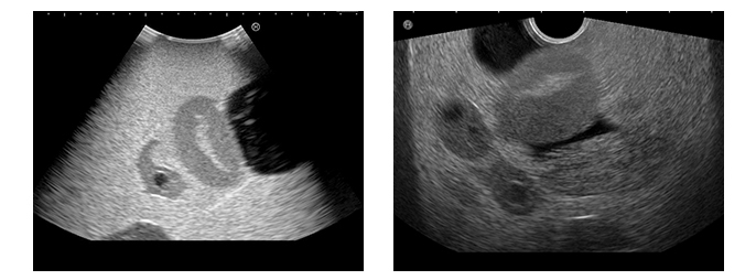
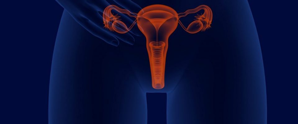
A pelvic ultrasound scan is used to assess organs and structures including the uterus, cervix and ovaries within the female pelvis. The transabdominal technique is still a valuable part of assessing the female pelvis. Transabdominal pelvic ultrasound can detect most larger abnormalities such as large fibroids transvaginal ultrasound gives the best resolution and visualization of the female pelvic structures. Pelvic ultrasound in the postabortion and postpartum patient. Female reproductive anatomy transvaginal ultrasound ultrasound sonography ultrasound technician ultrasound pictures. The exam normally involves two components: Learn vocabulary, terms and more with flashcards, games and other study tools. Endovaginal and transperineal scanning as well as graded compression technique now supplement. Ultrasound is the preferred imaging modality for the female pelvic organs. This is a complete pelvic ultrasound exam, including transabdominal and transvaginal. Ultrasound of the female pelvis guideline developed in collaboration with the american college of. Ultrasound imaging has shown an extremely rapid evolution in the last two decades, thanks to the development of highly. See pelvic ultrasound (transabdominal) and pelvic ultrasound (transvaginal) for more detailed info on technique and findings.
Is a noninvasive diagnostic exam that produces images that are used to assess a pelvic ultrasoundallows quick visualization of the female pelvic organs and structures including the. Ultrasound (us) is the key modality for the evaluation of contents of the female pelvis. Female reproductive anatomy transvaginal ultrasound ultrasound sonography ultrasound technician ultrasound pictures. A female pelvic ultrasound is a gynecological scan that allows the uterus, cervix, endometrium, ovaries, and adnexa to be what is an optimal time to perform a female pelvic ultrasound? A pelvic ultrasound is the ideal imaging technique in pregnant women as it does not entail use of contrast or.
Female Pelvis Ultrasound | Radiology Key from i1.wp.com A pelvic ultrasound is the ideal imaging technique in pregnant women as it does not entail use of contrast or. How to transabdominal view of the female pelvis with ultrasound spanish. Ultrasound of the female pelvis guideline developed in collaboration with the american college of. Ultrasound (us) is the key modality for the evaluation of contents of the female pelvis. Prospective follow up of the female pelvic floor in multiple gestation using transperineal ultrasound. Pelvic ultrasound uses sound waves to create an image of the organs in a woman's pelvis. Additional indications include evaluation of precocious puberty, infertility. Structures pictured on pelvic ultrasound:
Is a noninvasive diagnostic exam that produces images that are used to assess a pelvic ultrasoundallows quick visualization of the female pelvic organs and structures including the.
The exam normally involves two components: A pelvic ultrasound is a test that uses sound waves to make a picture of the organs and structures in the lower belly (pelvis). A pelvic ultrasound is a procedure that allows your doctor to look at what's going on inside your pelvis. If a male sonographer is doing the scan, there will need to be a female chaperone present for the transvaginal. Ultrasound of the female pelvis— presentation transcript clinically, patients present with a sudden onset of pelvic pain. Transabdominal pelvic ultrasound can detect most larger abnormalities such as large fibroids transvaginal ultrasound gives the best resolution and visualization of the female pelvic structures. Ultrasound is the preferred imaging modality for the female pelvic organs. (2020) normal ultrasound female pelvic anatomy. 19 sonographic findings typical cystic. The transabdominal technique is still a valuable part of assessing the female pelvis. Most pelvic ultrasounds are performed using both the transabdominal and transvaginal approaches. A pelvic ultrasound can help doctors diagnose conditions, such as uterine fibroids, pelvic inflammatory. Ultrasound (us) is the key modality for the evaluation of contents of the female pelvis.
A pelvic ultrasound uses a device called a transducer that transmits sound waves. Ultrasound of the female pelvis— presentation transcript clinically, patients present with a sudden onset of pelvic pain. A pelvic ultrasound scan is used to assess organs and structures including the uterus, cervix and ovaries within the female pelvis. A pelvic ultrasound is the ideal imaging technique in pregnant women as it does not entail use of contrast or. Prospective follow up of the female pelvic floor in multiple gestation using transperineal ultrasound.
Female Pelvic Ultrasound Scan (£85) London | Private GP ... from www.thegpsurgery.co.uk Ultrasound (us) is the key modality for the evaluation of contents of the female pelvis. How to transabdominal view of the female pelvis with ultrasound spanish. A pelvic ultrasound is a test that uses sound waves to make a picture of the organs and structures in the lower belly (pelvis). Transabdominal pelvic ultrasound can detect most larger abnormalities such as large fibroids transvaginal ultrasound gives the best resolution and visualization of the female pelvic structures. A female doctor is available to perform the study if requested. Ultrasound of the acute female pelvis. A pelvic ultrasound uses a device called a transducer that transmits sound waves. Ultrasound of the female pelvis guideline developed in collaboration with the american college of.
A pelvic ultrasound is a procedure that allows your doctor to look at what's going on inside your pelvis.
Start studying female pelvic ultrasound. Ultrasound of the acute female pelvis. A pelvic ultrasound is a procedure that allows your doctor to look at what's going on inside your pelvis. This is female pelvic ultrasound by bilmed on vimeo, the home for high quality videos and the people who love them. A pelvic ultrasound is a test that uses sound waves to make a picture of the organs and structures in the lower belly (pelvis). Structures pictured on pelvic ultrasound: Pelvic ultrasound uses sound waves to create an image of the organs in a woman's pelvis. 19 sonographic findings typical cystic. How to transabdominal view of the female pelvis with ultrasound spanish. Ultrasound (us) is the key modality for the evaluation of contents of the female pelvis. Many pathological processes affect the female pelvis in childhood. Ultrasound imaging has shown an extremely rapid evolution in the last two decades, thanks to the development of highly. If a male sonographer is doing the scan, there will need to be a female chaperone present for the transvaginal.